Indian Scientists Develop Advanced Brainstem Atlas
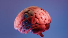
For many years, neuroscientists have been exploring the human brain similar to how early mapmakers charted unknown territories. Despite ongoing efforts, much of the brain's complex landscape remains uncharted, with pathologists typically examining only a few tissue samples from an organ that houses approximately 86 billion neurons. This is why the recent work by researchers at the Sudha Gopalakrishnan Brain Centre (SGBC) at the Indian Institute of Technology, Madras (IIT-M) is significant. They have developed what they claim is the most detailed three-dimensional atlas of the human brainstem, providing a cellular-level view.
Named Anchor (Atlas of Neurochemical Characterisation of the Human Brainstem with 3D Reconstruction), this digital map allows scientists to transition smoothly from MRI scans of the entire brain to individual nerve cells. It encompasses over 500 tissue sections from fetal, childhood, and adult brains, constructed using high-resolution microscope images instead of more expensive molecular methods. The result is a detailed map of the brainstem, identifying over 200 clusters of brain cells and nerve pathways.
The Crucial Role of the Brainstem
The brainstem, though a small part of the brain, is crucial for survival as it connects the brain to the spinal cord and manages essential functions such as breathing, heart rate, sleep, and movement. Damage to even small clusters of cells in this densely packed region can have severe consequences. Anchor's value lies not only in its detailed anatomical mapping but also in bridging the gap between medical imaging and cellular pathology.
As Shubha Tole, a neuroscientist at the Tata Institute of Fundamental Research, describes it, Anchor is an "unprecedented integration" of engineering, neuroscience, and medicine. This project positions India prominently on the global stage in neuroscience research.
Implications for Medical Research and Practice
Typically, neuropathologists begin by examining the entire brain post-mortem or during neurosurgery, looking for clues with the naked eye before delving into microscopic analysis. However, modern MRI scans, while comprehensive, lack cellular detail, and microscopes can only reveal isolated slices of cells. The Anchor atlas aims to close this gap, enabling users to zoom in from the entire brainstem to individual neurons while maintaining spatial accuracy.
The atlas is freely available online and is expected to serve as a valuable reference for neuroscientists, neurologists, and neurosurgeons worldwide. Its applications extend beyond anatomy, potentially aiding in the understanding of disorders such as Alzheimer's, Parkinson's, stroke, and sudden infant death syndrome (SIDS), and enhancing surgical precision in the brainstem.
"Anchor's greatest value lies in the questions it could help answer," says Partha Mitra, a brain scientist at Cold Spring Harbor Laboratory.
By comparing healthy brainstem maps with diseased ones, scientists might gain insights into how diseases alter brain structures at the cellular level. The atlas could also help explain how infections, including Covid-19, cause long-term neurological damage.
Future Directions and Potential
The SGBC plans to expand this work by imaging over 100 whole human brains across various life stages and neurological conditions, creating a comprehensive reference library. While the atlas won't solve all the mysteries of the human brain, it provides a more detailed map, enabling scientists to pose more informed questions.
Every brain, as noted by Rebecca Folkerth, a neuropathologist affiliated with Harvard Medical School, is a "treasure chest of new knowledge." This project, with its combination of high-resolution imaging and detailed anatomical mapping, represents a significant advancement in the field of neuroscience.
Source: Original Article